For people living with multiple sclerosis (MS), understanding how the disease progresses and how well treatments are working has traditionally relied on tools like MRI scans and clinical assessments. While useful, these tools have their limitations: they are expensive, not always readily accessible, and can miss subtle or early signs of disease activity. A recent study by Kuhle et al. (2019) brings a promising biomarker into the spotlight—neurofilament light chain (NfL)—a protein found in blood that could change how we monitor and manage MS.
What Is Neurofilament Light Chain?
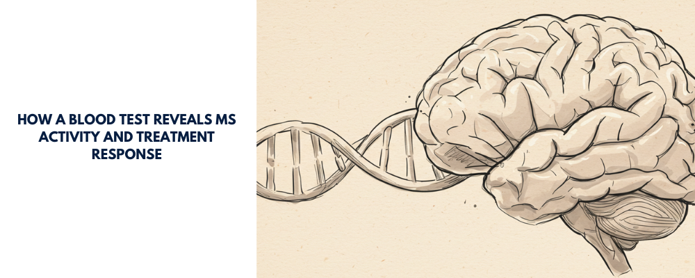
Neurofilament light chain is a structural protein found in neurons. When neurons are damaged—a hallmark of MS—NfL leaks into the cerebrospinal fluid (CSF) and eventually into the bloodstream. Technological advances, particularly the SIMOA assay (Single Molecule Array), have made it possible to detect tiny amounts of NfL in blood samples with high sensitivity. This paves the way for a minimally invasive method to track neurodegeneration in real-time.
The Study: Two Major Clinical Trials
The study by Kuhle and colleagues analyzed blood samples from 589 patients with relapsing-remitting MS (RRMS) participating in two Phase 3 trials:
FREEDOMS: A two-year, placebo-controlled trial of the oral drug fingolimod.
TRANSFORMS: A one-year, active-controlled trial comparing fingolimod to interferon beta-1a (IFN-β-1a).
The research also included samples from 35 healthy controls for comparison.
Key Findings
1. NfL Levels Reflect MS Disease Activity
MS patients had significantly higher NfL levels than healthy individuals (30.5 pg/mL vs 16.9 pg/mL).
Higher NfL levels correlated with MRI findings, such as:
T2 lesion volume (reflecting total disease burden).
Gadolinium-enhancing lesions (indicating active inflammation).
Even patients without visible lesions on MRI had elevated NfL levels, suggesting the marker detects damage beyond what imaging can show.
2. Baseline NfL Predicts Future Disease Course
Patients with high NfL levels at baseline were more likely to experience:
More new or enlarging lesions.
Higher relapse rates.
Greater brain volume loss.
A trend toward increased risk of disability worsening.
3. Fingolimod Effectively Lowers NfL
Treatment with fingolimod significantly reduced NfL levels within 6 months.
This reduction was sustained over the study duration and approached the levels seen in healthy individuals.
Interferon also reduced NfL levels, but less dramatically than fingolimod.
Why This Matters
NfL in blood could become a game-changing biomarker for MS. It offers a fast, inexpensive, and repeatable method to assess:
Ongoing neural damage.
Response to treatment.
Risk of future disability.
Unlike MRI, which requires costly equipment and can miss invisible damage, a blood test can offer real-time insights into disease activity.
Limitations and Future Directions
While the study shows promising results, it’s important to note:
This was a post hoc analysis using data from already completed trials.
More work is needed to establish reference ranges and account for factors like age and other neurological conditions.
The clinical utility of NfL for individual treatment decisions still needs validation.
Conclusion
Blood NfL is emerging as a powerful tool in the fight against MS. It provides a window into the invisible processes of neurodegeneration and inflammation. With further validation, it may soon join the clinical toolkit to help neurologists make smarter, faster, and more personalized decisions for their patients.
Disclaimer: This blog post is based on the information provided in the cited scientific article. It aims to provide an accessible summary of the research findings and should not be considered as definitive medical advice. For any health concerns, please consult with a qualified healthcare professional.
Reference:
Kuhle J, et al. Blood neurofilament light chain as a biomarker of MS disease activity and treatment response. Neurology. 2019;92:e1007–e1015.